Dr. Randi Fredricks, Ph.D.
 Mood and anxiety disorders are characterized by a variety of neuroendocrine, neurotransmitter, and neuroanatomical disruptions. Identifying the most functionally relevant differences is complicated by the high degree of interconnectivity between neurotransmitter- and neuropeptide-containing circuits in limbic, brain stem, and higher cortical brain areas.
This alteration in brain structure or function and in neurotransmitter signaling may result from environmental experiences and underlying genetic predisposition; such alterations can increase the risk for psychopathology.
The Brain, Fear and Anxiety
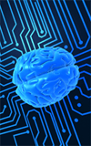
There are several parts of the brain that act in concert in a highly dynamic interplay that gives rise to fear and anxiety. By using brain imaging technologies and neurochemical techniques, scientists have discovered that a network of interacting structures is responsible for these emotions.
Researchers involved in anxiety treatment have found that there are several parts of the brain play a role in fear and anxiety. Using brain imaging technologies and neurochemical techniques, scientists are finding that a network of interacting structures is responsible for these emotions. Most of this research centers on the amygdala, an almond-shaped structure deep within the brain.
The amygdala serves as a communications hub between the parts of the brain that process incoming sensory signals and the parts that interpret them. It signals that a threat is present, and triggers a fear response or anxiety. Emotional memories stored in the central part of the amygdala play a role in a variety of anxiety disorders.
Research Into Anxiety and the Brain
Research into anxiety and the brain has also examined the hippocampus, another part of the brain responsible for processing threatening or traumatic stimuli. Part of the hippocampus’ role is to encode information into memories. Studies have revealed that the hippocampus tends to be smaller in people who have undergone severe stress because of child abuse or military combat. Researchers believe that this reduced size inhibits the hippocampus from properly processing traumatic events.
Other scientists have determined that the basal ganglia and striatum in the brain are involved in specific anxiety disorders, like OCD. People with anxiety suffer from is a heightened autonomic nervous system (ANS) reaction to a perceived threat.
One the main treatment goals in therapy for anxiety disorders is to use specific methods to diminish the ANS reaction, eliminate the anxiety symptoms, decrease the cycle of avoidance, and improve overall well-being.
Anxiety disorders often become so severe that normal life and relationships become impaired. There are many types of anxiety disorders with their own unique sets of symptoms. Some of these disorders include panic disorder, obsessive-compulsive disorder (OCD), post-traumatic stress disorder (PTSD), social phobia (or social anxiety disorder), specific phobias, and generalized anxiety disorder (GAD).
By learning more about brain circuitry involved in fear and anxiety, researchers will be able to devise new and more specific treatments and counseling approaches for anxiety disorders. For example, it someday may be possible to increase the influence of the thinking parts of the brain on the amygdala, placing the fear and anxiety response under conscious control. Additionally, neurogenesis (birth of new brain cells) throughout life may be the key to developing a a method to stimulate growth of new neurons in the hippocampus in people with severe anxiety.
References
Egashira N, Tanoue A, Matsuda T, et al. Impaired social interaction and reduced anxiety-related behavior in vasopressin V1a receptor knockout mice. Behav Brain Res. 2007;178:123–127.
Kendler KS, Gardner CO, Lichtenstein P. A developmental twin study of symptoms of anxiety and depression: evidence for genetic innovation and attenuation. Psychol Med. 2008;38:1567–1575.
Hallett V, Ronald A, Rijsdijk F, et al. Phenotypic and genetic differentiation of anxiety-related behaviors in middle childhood. Depress Anxiety. 2009;26:316–324.
Lee YS, Hwang J, Kim SJ, et al. Decreased blood flow of temporal regions of the brain in subjects with panic disorder. J Psychiatr Res. 2006;40:528–534.
Engel K, Bandelow B, Gruber O, et al. Neuroimaging in anxiety disorders. J Neural Transm. 2009;116:703–716.
About the Author
Randi Fredricks, Ph.D. is a practicing therapist, researcher and author specializing in the treatment of anxiety, depression, addiction, eating disorders, and related disorders. Dr. Fredricks is a best-selling author of numerous books on complementary and alternative treatments for mental health including Complementary and Alternative Treatments for Anxiety. For more information on Dr. Fredricks work, visit her practice website San Jose Counseling and Psychotherapy.